— Vernon R. Stanley, MD, PhD ©2019. All rights Reserved.
In the blog post below, we will discuss the finding of “ST-segment elevation” and a few of the differential diagnosis to keep in mind as this observation is made on the ECG. Please understand these are only a handful of differential diagnosis as regards ST-segment elevation; however for the sake of brevity we will narrow the discussion to comparison of the following, namely in this post we will discuss:
Pericarditis- Benign Normal Variant
- Acute Anterior STEMI
Review from Part 1: Acute Pericarditis – click here.
Early Repolarization Example…
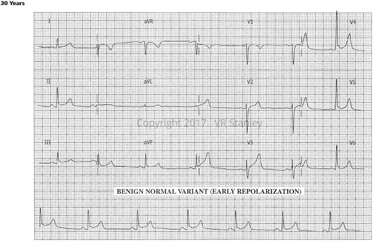
Benign Normal Variant (Early Repolarization)
During your daily practice of medicine, you are frequently faced with the following scenario:
You are presented with a cardiogram which demonstrates minor, subtle ST-segment elevations concave up. After analyzing the tracing, you have narrowed your choices to:
- Benign Normal Variant (Early Repolarization).
- Early, suspected Acute STEMI.
The sobering ramifications of this decision-making are obvious. On the surface, this challenge might appear to be straightforward; but in the clinical world you will encounter tracings that challenge even the most expert eye.
General comments regarding Early Repolarization Pattern
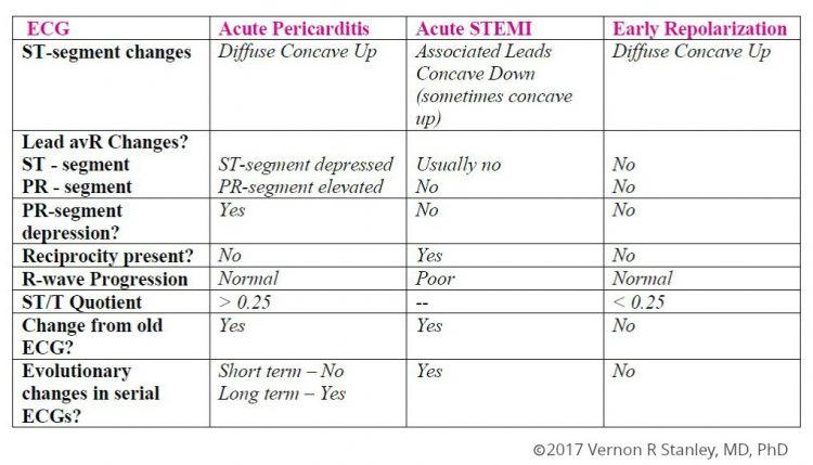
- Serial tracings will show essentially no changes evolving (unless an infarction is evolving also)
- Early Repolarization patterns essentially never show “terminal QRS distortion” (further discussion in our post on Terminal QRS Distortion)
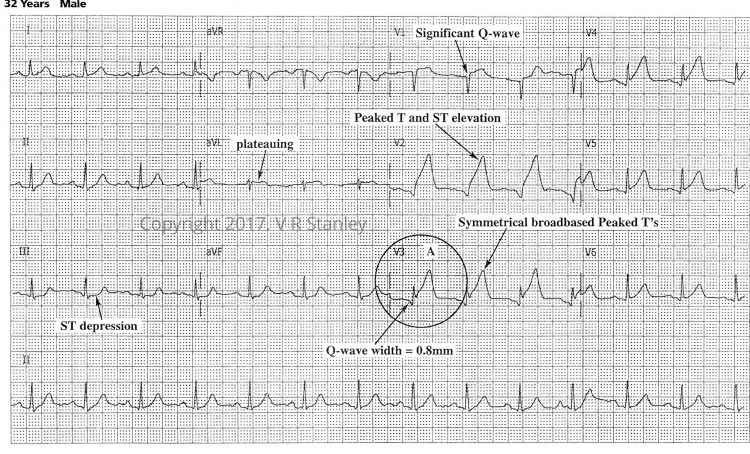
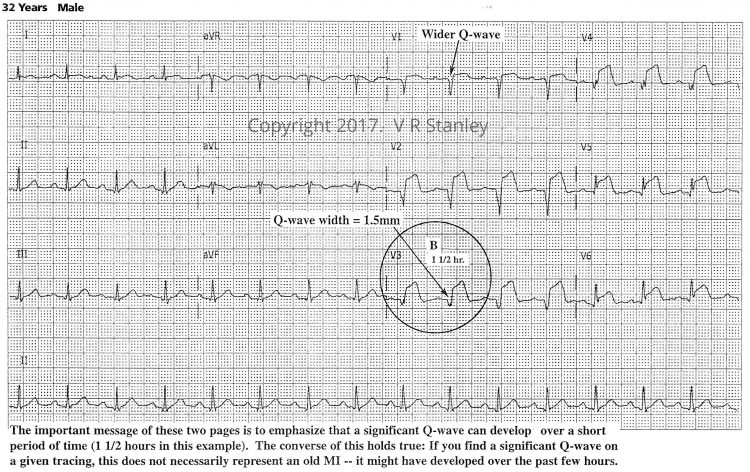
Let us now review two serial tracings of an Acute Anterolateral STEMI – tracing “A” and tracing “B” at 1.5 hours.
TRACING A: The tracing below represents the initial presenting cardiogram of a 32 year old with ACS.
The tracing below is a serial tracing of the same patient at 1.5 hours later…
I want you to focus on two aspects of these tracings:
- The low point (nadir) of the S-wave of Leads V1, V2, V3
- The terminal R-wave of Leads V4, V5, V6
FINDINGS SUGGESTIVE OF ACUTE ANTERIOR STEMI (as opposed to benign normal variant)
- ST-segment elevation, concave down (tombstoning, plateauing; sometimes concave up) in Leads V1 to V4 (maximum Leads V2, V3)
- ST-segment depression of reciprocity (especially in Leads II, III, avF, V5, V6)
- Poor R-wave progression
- Anterior Q-waves present
- Late transition
- Presence of Hyperacute Ts
- Terminal QRS distortion
Please be aware that sometimes the ST-segment elevation is concave up (even in the setting of the acute STEMI). In these cases it is especially important to carefully look at all the clues, especially the “terminal QRS distortion”.
To summarize….
Questions? Contact Us


2 Responses
My husband had an emergency cath last night because of this.
Hello Ms Meyer, We are sorry to hear this and hope your husband is doing better today. We send our regards and prayers for full and fast recovery. Sincerely, ECGcourse Support Team