Adult Long Bones Anatomy Copy

The long bones comprise a large group of bones including the– humerus ,clavicle, radius, ulna, metacarpals, phalanges of feet and hands, femur, tibia, fibula, metatarsals. The common landmarks and anatomic parts of the adult long bones are shown in Illustration 41 below.

A common challenge in your practice of medicine will be to evaluate the patients with trauma to the long bones. This Module will begin the discussion of trauma to the Long Bones with the goal of determining whether a fracture is present, describe the fracture often with the end result of presenting to a receiving practitioner ( often an orthopedic surgeon ). In doing so we may discuss a treatment plan.


ill51redo-750x1187


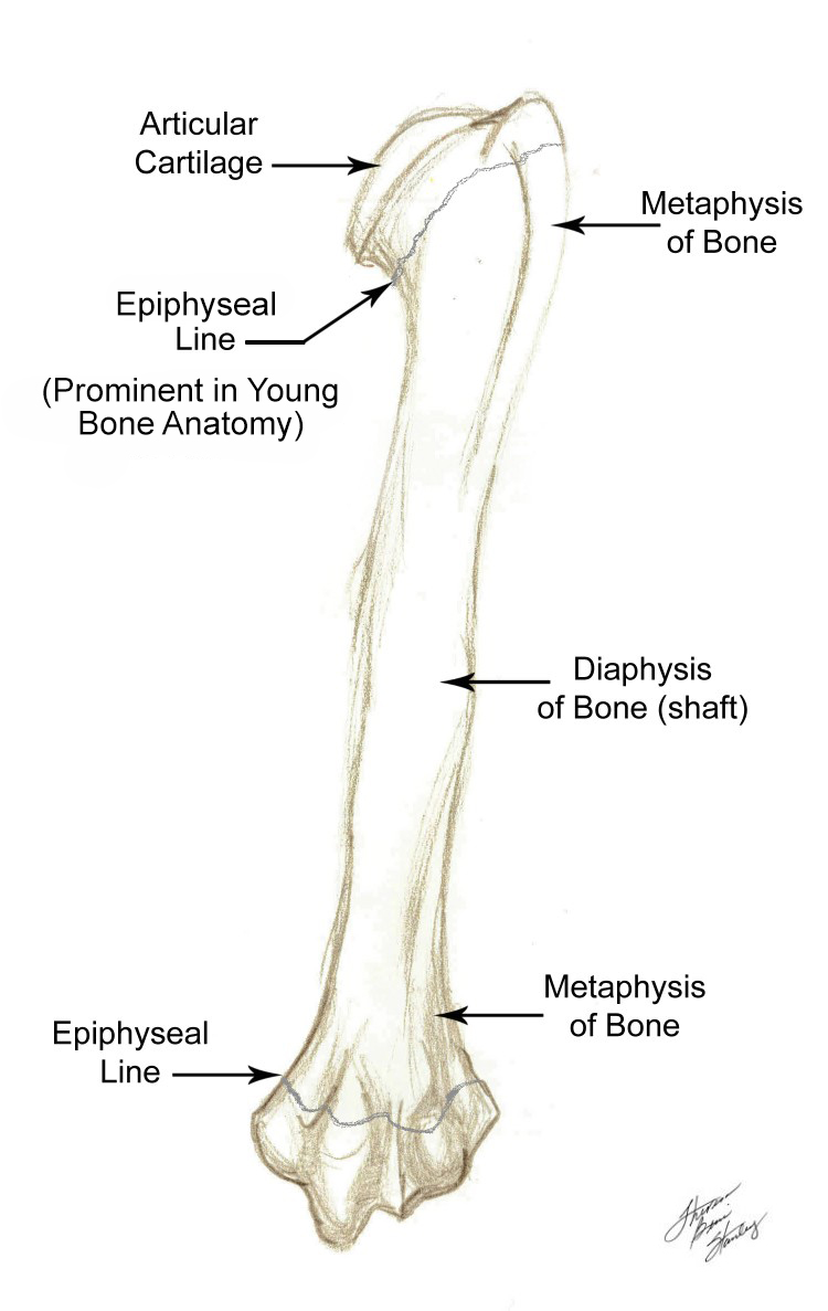
Illustration 41 Adult Long Bones Anatomy

. . . First of all, the fractures are described as open or closed.:

OPEN
fracture associated with a laceration or puncture wound communicating with the bone fracture.

CLOSED
no laceration or puncture wound communicating with the bone fracture.

Secondly we describe the fracture as to location, severity of the fracture including fragmentation, separation, angulation, as well as neurological, muscular functional deficiency and vascular status. Let us begin by presenting a series of artist’s illustrations of the most common types of fractures you will encounter in your practice. These will be presented in Illustrations 52A thru 52U.

To begin this series

click on

NEXT