. . . …………………………….. COLOR CODE GUIDE …………………………..
X-Ray examinations (plain film and CT) are generated by an X-Ray beam passing through the patient (or body part). The X-Ray beam is partially absorbed (or reflected) proportionate to the body part radio-density with a small percentage of the radiation exiting the patient and striking the detector (film) where the image is digitized and stored as a hard copy film, viewed on a monitor screen or transmitted to a remote location for viewing. The images will appear as black and white and infinite shades in between.
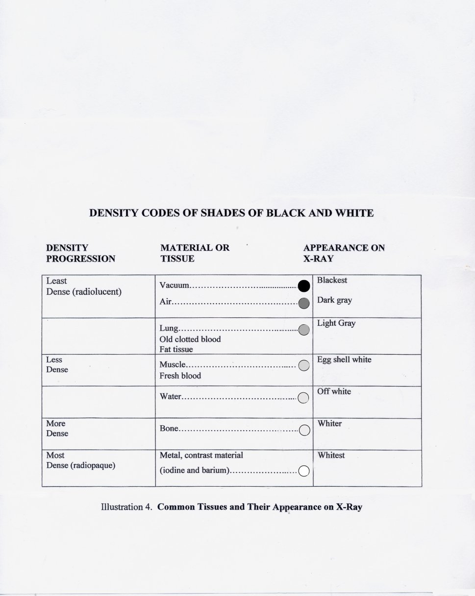
Another important characteristic is that of homogeneous or heterogeneous, e.g., air would be homogeneous black, piece of metal homogeneous white , lung tissue would be heterogeneous (speckled black and white), etc. A guideline for the interpretation of these shades is given in Illustration 4. With this color code information, we can clearly predict how a chest X-Ray will appear, e.g.,
…..the lung field will be “dark gray speckled with white.”
…..the heart image will be “egg shell white.”
….the ribs will appear “white.”
…..the hemi-diaphragm will appear “egg shell white.”
…..calcifications will be “white.”
…..air pocket would be homogeneous black…..etc.
With this predicted color coding, we now simply fill in the shape of e.g., lung, heart, ribs, diaphragm, fresh blood, old blood etc. this systematic prediction should be applied to all other X-Rays or body parts (e.g., knee wrist, abdomen, etc. ) following a similar conclusion of predicted color shades and shapes.
The converse clearly holds true:
As you analyze an X-Ray you will note multiple areas of black–gray–off white–white, each shade associated with a shape. Your job as the interpreter is to label each area as e.g., fresh blood, heart, lung, fat, bone, muscle, fluid, pneumonia, effusion, air etc.
for a PEARL summary of MODULE I
click on
” NEXT ”