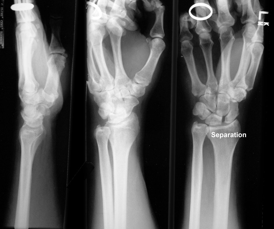
. . Note the increased spacing between the scaphoid and lunate. It is curious to note that on the lateral view the carpal bones (lunate, capitate ) are properly aligned with the radius axis ——–i.e. there is no lunate or perilunate dislocation. Please scroll back and note the increased scapholunate spacing of Illustration 78——in the absence of the lateral view I am unable to determine if there is a lunate dislocation, perilunate dislocation or neither. NOTE These are beautiful examples to demonstrate the need to look at all three views since the abnormality may be revealed in only one view. Your systematic analysis of X-Ray and CT interpretation must include looking at all views as you search for pneumonia, pneumothorax, masses, fractures, dislocations, air, misalignments, acute and old bleeding, etc