The purpose of this module is to address the issue of injuries to the upper extremity. We will begin with the shoulder and work our way down to the hand. We will present examples of common fractures and dislocations to each area and where appropriate will briefly discuss clinical aspects such as mechanism of injury and treatment pearls. Each presentation will be in the form of an actual radiograph and/or a medical illustration sketch with a label and sometimes color-coding identifying the salient feature. I again remind you to utilize the built-in PAC system —especially zooming in and hot-lighting the area of interest.
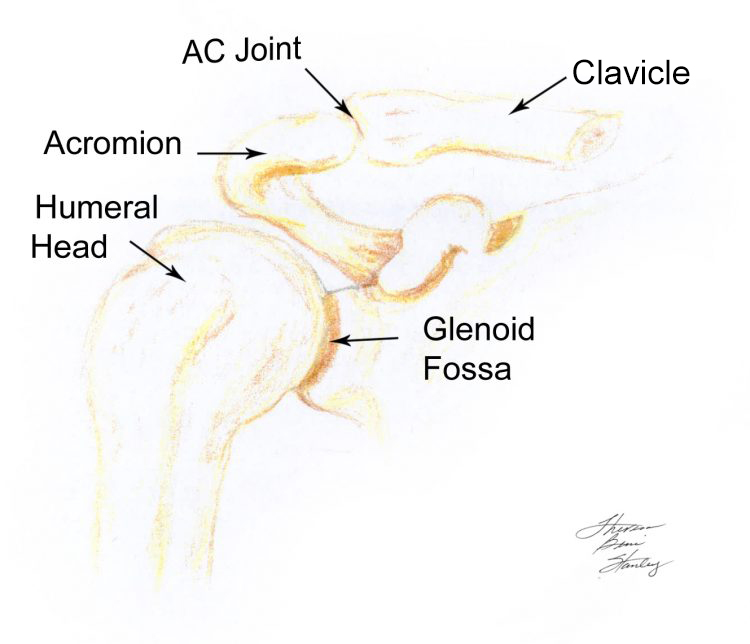
Illustration 45 Shoulder Anatomy
. . . See Illustration 45 for an artist sketch of a normal shoulder and Illustration 46 for a radiograph of a normal shoulder .

