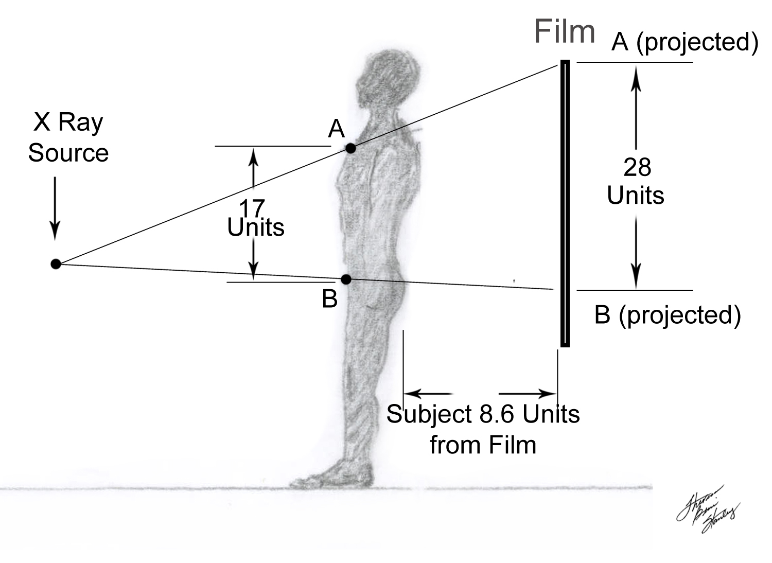
Illustration 3 B Source to Film—- fixed distance
CONCLUSION——– The closer the subject is to the film —————-the smaller, more true to form and sharper focus will be the image. .
. . . . . . We know that the X-Ray image
is black and white ( and all the infinite shades of gray in between ). Our challenge is to focus on a given area of the X-Ray and match it according to location, color [ white….light gray…..dark gray………black ] and shape with a differential diagnosis of :
normal,
bone,
tumor,
pneumonia,
blood,
muscle,
air,
lung,
fat,
etc.
The task is aided and the differential narrowed by clinical correlation.
Let us now address the question of color coding of common tissues.
Please click on
” NEXT “