(C) Revised 2024 ECGcourse.com, LLC. All rights reserved. | Author: Vernon R Stanley, MD, PhD | Editor: Courtney Stanley, MSPA-C
Please answer the following questions. (Answers at bottom of blog.)
Q1: During the evolution of an Acute MI, you might see:
- a. ST-elevation concave down
- b. Development of significant Q-wave
- c. T-wave inversion
- d. Reciprocal ST-depression
- e. All the above
Q2: Sometimes in the setting of an Acute MI, you will see ST ___________________ in the Associated leads and ST _____________________ in the Reciprocal Leads. This is virtually diagnostic of the Acute MI in the absence of LBBB and LVH.
- a. depression, elevation (concave down)
- b. elevation (concave down), depression
- c. asymmetry, sloping
- d. sloping, asymmetry
You are working as a mid-level practitioner at an urgent care clinic in rural Mt Juliet, Tennessee, USA. The patient below presents with jaw and left shoulder pain. Your interpretation and disposition is:

INTERPRETATION:
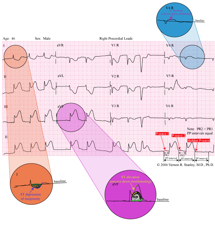
- The underlying ECG rhythm is second degree AV Block Mobitz I (Wenckebach) 3:2. The ECG rhythm per se will usually require no treatment
- Acute Inferior-Right Ventricular STEMI as manifested by ST elevation concave down in the associated leads with ST-depressions in the reciprocal leads. Administer your protocol meds including thrombolytics (if no contraindications) or to the cath lab. Please note that the ST elevation shape takes on the eerie appearance of a tombstone, then the term “tombstoning ECG“.
FOCUS AND GOAL OF THIS LESSON:
- Review of characteristics of the Acute ST elevation MI (in particular, we will analyze a tracing of the acute inferior MI and investigate the question of the acute right ventricular MI). A discussion of the tombstoning ECG will be intrinsic to this blog post.
- Recognition of Second-Degree AV Block both Mobitz I and Mobitz II.
DISCUSSION:
What is a Tombstoning ECG and Why Does it Matter?
In the practice of medicine, we often hear the term Tombstoning ECG. But what does it reference generally and why does it matter? The term tombstoning is used to reference the eerie appearance of the ST segment elevation seen in the 12-lead ECG of a patient having an Acute ST Elevation MI (aka STEMI). When present, it may be associated with more myocardial damage, lowered right ventricular function and a poorer patient prognosis (NIH 2009 B. Bahattin).
Because time is cardiac muscle, it is critical that each member of the medical team be aware of this visual phenomenon of the tombstoning ECG and continue to look for such changes on the 12-lead in patients presenting with Acute Chest Pain or a history consistent with Acute Coronary Syndrome. Excellent examples of Acute ST Elevation MIs (some with tombstoning) may be seen here as well: Anterior Myocardial Infarction • LITFL • ECG Library Diagnosis
Additional notes on this ECG Rhythm…
Recommended additional reading on Mobitz 1 vs 2: Rhythms Review: Is this Second Degree AV Block 2:1 Mobitz I or Mobitz II? – ECGcourse.com
- SECOND DEGREE AV BLOCK
The second degree AV Block falls into two distinct categories. Remember that the characteristic of normal sinus rhythm is that every P-wave is followed by a QRS complex, i.e. the ratio of P:QRS is 1:1. If this ratio is not 1:1 but is 2:1, 3:1, 3:2, 4:3…… and there IS a relationship between the P and the QRS ( ie this excludes the complete heart block), then the rhythm falls into the category of second degree AV block.
There are two distinct types distinguished by the relationship between the P-wave and the QRS complex, i.e. the PR- interval pattern. They are as follows:
A. MOBITZ I (Wenckebach)
ELECTRICAL CHARACTERISTIC: The signal begins in the SA node, travels to the AV node, then to the ventricles. This produces a normal P-QRS-T complex. The next signal from above again begins in the SA node, travels to the AV node and travels more slowly across the AV node producing a longer PR interval. The PR interval progressively gets longer until a beat is completely dropped. The cycle then repeats itself. The ratios of P:QRS will occur as 2:1, 3:2, 4:3, 5:4, 6:5, 7:6, etc.
Please scroll up and note the labels of the rhythm strip with a ratio of 3:2 = P : QRS noted.
Wenckebach is usually a benign rhythm and most often requires no treatment. It occasionally occurs during the evolution of the MI, is often transient and self-limiting.
MOBITZ I (Wenckebach) is CHARACTERIZED BY The PR interval getting progressively longer until one P-wave is blocked. The cycle then starts again.
B. MOBITZ II SECOND DEGREE AV BLOCK
ELECTRICAL CHARACTERISTIC: Malfunctioning AV node (or commonly more distal conduction tissue) of Mobitz II is characterized as follows: if the P-wave is conducted, the PR interval is constant. Otherwise, the P-wave is not conducted. Mobitz II is a more threatening rhythm than Mobitz I (Wenckebach) and is sometimes followed by complete heart block. ACLS protocol recommends to prepare for possible external pacemaker.
MOBITZ II CHARACTERIZED BY the PR intervals on conducted beats are equal, some of the P-waves are blocked.
THE ACUTE ST-ELEVATION MI (ACUTE MI; STEMI)
Now scroll up and examine this 12-lead ECG Case Study tracing. Note that I have circled and magnified two Leads I and avF. The ST elevation of Lead avF (as well as II and III) takes on the shape concave down. This particular cardiogram demonstrates ST elevation resembling the edge of a tombstone (aka tombstoning ECG). Furthermore, note the ST depressions in Leads I, avL, V1-V6. Take note of the artist’s rendering the tombstone with its edge blending in with the curvature of the ST segment. An additional finding, which virtually clinches the diagnosis, is the finding of reciprocity, i.e. the leads opposite the associated leads will be depressed. In this case the opposite (reciprocal) leads are I, avL, V1-V6. This concept is emphasized by the artist’s rendering of a shallow grave with a casket, a reminder that when you see a Tombstoning ECG, it is indeed and ominous sign which needs great attention from the provider.
Please know that the patient with the acute MI does not always exhibit changes so obvious as this one. In fact, reciprocal depressions are often absent; and the ST elevations may not be as dramatic as the tombstoning ECG demonstrated here, but the ST depressions are indeed usually concave down.
WARNING about Tombstoning ECG mimics or distortions:
Don’t forget about the two greatest booby traps of electrocardiography: the LBBB and the LVH pattern.
Both these patterns are fraught with ST-T-Q waves that are often pseudoinfarction and pseudoischemia changes.
ACUTE RIGHT VENTRICULAR INFARCTION
It is interesting to note that no Lead of the standard 12-Lead “looks at” the right ventricle or the posterior aspect of the heart and it is therefore true that we can only obtain indirect information about these portions of the heart. It is extremely difficult if not impossible to make the diagnosis with certainty of the acute right ventricular or acute posterior infarction unless you place special leads on the chest.
In order to monitor the right ventricle, one must place special leads on the chest as follows:
The right precordial Leads V1R, V2R, V3R, V4R, V5R, V6R
If one looks at the right precordial Leads of the normal heart, you will note that the ST segments will lie on the baseline.
If you now examine the right precordial leads in the patient experiencing an acute RV infarction, you will often find ST elevation concave down, especially seen in Lead V4R————This tracing shows exactly that and represents an acute RV MI.
PEARL: The Acute Right Ventricular infarction and Acute Posterior Infarctions are frequently associated with the Acute Inferior MI
SUMMARY RECOMMENDATION WHEN LOOKING FOR THE ACUTE RV INFARCTION
In the setting of an Acute Inferior MI, you should “think” RV infarction and proceed to look for it. This search will necessitate the placement of the right precordial Leads (V1R, V2R, V6R).
The primary focus should be on Lead V4R since it is the best lead that “looks at” the right ventricle and indeed here lies the diagnosis:
Acute Right Ventricular Infarction – ST segment elevation (concave Down) in Lead V4R
INTERPRETATION:
- Tombstoning on the ECG is noted here. Changes are consistent with the Acute Inferior-Right Ventricular STEMI as manifested by ST elevation concave down in the associated leads with ST-depressions in the reciprocal leads. Administer your protocol meds including thrombolytics (if no contraindications) or send patient to cath lab.
- The underlying rhythm is second degree AV Block Mobitz I (Wenckebach) 3:2. The rhythm per se will usually require no treatment
Q1: During the evolution of an Acute MI, you might see:
- a. ST-elevation concave down
- b. Development of significant Q-wave
- c. T-wave inversion
- d. Reciprocal ST-depression
- e. All the above
Q2: Sometimes in the setting of an Acute MI, you will see ST ___________________ in the Associated leads and ST _____________________ in the Reciprocal Leads. This is virtually diagnostic of the Acute MI in the absence of LBBB and LVH.
- a. depression, elevation (concave down)
- b. elevation (concave down), depression
- c. asymmetry, sloping
- d. sloping, asymmetry
Additional Reading on Tombstoning ECGs
- Dr Stanley’s 12-lead Textbook, Workbook & HEART Ruler Packet – ECGcourse.com
- Dr. Smith’s ECG Blog (hqmeded-ecg.blogspot.com)
- Marriott’s Practical Electrocardiography: Strauss MD PhD, David G., Schocken, Douglas D.: 9781496397454: Amazon.com: Books
Questions? Contact Us
Specials & Newsletter SignUp
Form for Specials Page.


1 Response