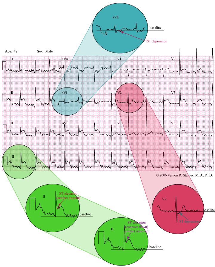
Posterior MI – the Hidden STEMI
©2020 Renewed. Vernon R Stanley, MD, PhD
INTERPRETATION:
- Early Transition.
- Suspicious of Acute Posterior MI (STEMI).
- Artifact.
- Acute inferolateral MI (STEMI).
THE POSTERIOR MI
The posterior aspect of the heart will of course be subject to ischemia and infarction (acute and old) as is any other area of the heart. The status of the posterior aspect is determined by analysis of the lead(s) that “looks at” the posterior aspect of the heart. No lead of the standard 12-lead looks directly at the posterior aspect of the heart. We must therefore rely on indirect measurements and consequently the information is of more limited value, unless we place special leads on the chest.
Please know that the posterior infarction is frequently associated with the acute inferior infarction. The clinical meaning is simply that in the setting of the Acute Inferior MI, you should “think” Acute Posterior Infarction and proceed to look for it. This statement also applies for the acute Right Ventricular Infarction.
We will agree that the precordial leads V1 and V2 indirectly view the posterior aspect of the heart (they are on the other side of Alice’s looking glass). If the posterior aspect is injured, it’s voltage contribution will be lowered and therefore from the vantage point of the anterior chest, the R-wave will heighten (since the anterior depolarization vector is opposed by the posterior depolarization vector). It then follows that a taller than usual R-wave in leads V1 and V2 will result in the case of the posterior infarction.
A direct monitor of the posterior aspect of the heart is reflected in special leads Vposterior by placing special electrodes on the patient’s back. These leads are designated V7-V10.
If we are looking for evidence of an acute posterior MI or ischemia, then we as per usual look for ST-elevations and/or T-wave inversions in leads V7-V10. Implicit in this presentation is that the posterior MI may result in early transition (due to losses of posterior forces in leads V1 and V2).
CONCLUSION:
Findings suggestive of acute POSTERIOR myocardial infarction:
A. ST-segment depression in Leads V1, V2.
B. Early transition.
C. ST-segment elevation concave down Leads V8,V9,V10
Therefore, in the setting of the acute inferior myocardial Infarction, you should then request that the posterior leads be placed. If analysis of these posterior leads reveals ST elevation concave down especially in Lead V8, V9, V10, this is virtually diagnostic of the Acute Posterior MI .
GENERAL RECOMMENDATION
When analyzing a tracing, you should carry a magnifying glass and magnify the images as I have demonstrated in this Case Study tracing. Mentally color-code areas of the waveform and activate your built-in human brain filter and remove artifact and smooth out the wandering baseline….
Now make your decision of interpretation as follows:
INTERPRETATION:
- Early Transition.
- Suspicious of Acute Posterior MI (STEMI).
- Artifact.
- Acute inferolateral MI (STEMI).


One Response