Before we begin to delve into the STEMI review course, let us first go back to basics to review the Normal ECG.
THIS SECTION CONTAINS:
- The concept of vectors will be revisited especially as it applies to the development of the waveshape of Lead V 1 and Lead V 6 .
- The following characteristics of the depolarization wavefront of the septum and the RV/LV will be the focus of the topic:
- The timeframe in which the depolarization wavefront occurs.
- The magnitude of the depolarization wavefront.
- The direction of the depolarization wavefront.
- The position of the electrode.
- Introduction of the Notational Standards of QRS-complex: P-QRS-T
- Intervals to be routinely measured— PR-Interval, QT-Interval, QRS Duration
- The concept of automaticity
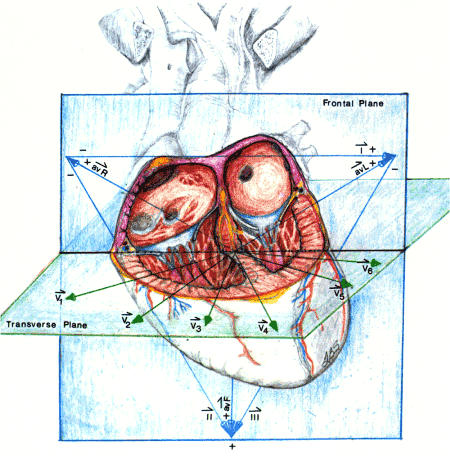
The purpose of each vector is to orient the student regarding representation of each lead. Each lead reflects a vantage point voltage as viewed from one specific angle. Three scenarios again possible: 1. ECG voltage deflection positive if wavefront traveling down shaft of arrow toward point. 2. ECG voltage deflection negative if away from the point. 3. ECG voltage deflection zero if perpendicular to arrow shaft.
For further discussion of Vectors…..
The essence of the understanding and development of the waveshape patterns of the 12-Lead lies in the following principles:
- The ventricles are depolarized in a wavefront manner with definite DIRECTION and MAGNITUDE .
- The ventricles are depolarized in a definite time sequential manner dictated by the conduction system ( SA-Node,Bundle of HIS with all its branches and terminating in the Purkinje Cells ).
- The ECG equipment is designed to sample the heart voltage with particular focus on the DIRECTION andMAGNITUDE of the depolarization activity.

This illustration shows propagation of electrical signal from SA node to Purkinje cells. Notice the wavefront nature and direction of the depolarization signal. The propagation of the wavefront through the myocardium is myocardial cell to myocardial cell to myocardial cell etc., etc., with termination in the right side of septum and epicardium of R and L ventricle.
Note the following time notations:
- t 1 = time of initiation of depolarization of atria.
- t 2 = time of initiation of depolarization of the AV Node
- t 3 = time of initiation of depolarization of septum
- t 4R = time of initiation of depolarization of Right Ventricle
- t 4L = time of initiation of depolarization of Left Ventricle
For a discussion of the phenomena of Automaticity ——– click “MARK COMPLETE” or ” NEXT “