(C) 2025 ECGcourse.com LLC All rights reserved | Author Vernon R. Stanley, MD, PhD | Co-editor Courtney Stanley, MSPA-C |
When interpreting a LBBB or RBBB on ECG, it is very common that the computer interpretation on a 12-lead ECG will read “non-specific T-wave changes”. The computer interpretation on the ECG is notoriously unreliable when analyzing the T-wave and ST segment. The gamut of T-wave changes which are noted can be secondary to anything from a benign variant of normal to ominous, early changes of an acute MI.
Before we begin this discussion, we invite you to answer the following questions. (Answers are posted at the bottom of this post.)
Question #1: Differentiating between the LBBB pattern and the RBBB pattern is best accomplished by referring to Lead ___
A. Lead III
B. Lead I
C. Lead avR
D. Lead V1
Question #2: In an otherwise normal case of LBBB & RBBB, if the terminal portion of the QRS complex is positive, the T-wave will be inverted. This is described as ________ T-wave changes.
A. Primary T-wave Changes (or Concordance of the T-waves)
B. Secondary T-wave Changes (or Discordance of the T-waves)
C. Tertiary T-wave Changes (or Accordion Effect of the T-waves)
Question #3: The required |QRS| > 0.12 seconds is common to both the RBBB and the LBBB.
A. True
B. False
Classic LBBB on 12-lead ECG
The finding of QRS = 0.12 sec should prompt you to proceed to look for the following potential scenarios:
1. LBBB
2. RBBB
3. IVCD (IntraVentricular Conduction Delay)
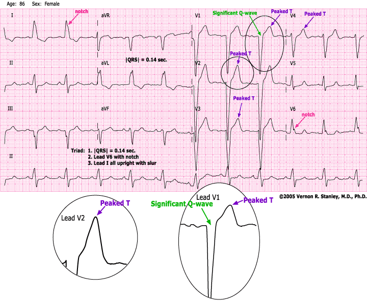
You will note that this ECG tracing satisfies the Classic Triad of the LBBB as follows:
1. |QRS| > or = 0.12 sec
2. RSR’ (slurred or notched) in Leads V6, V5 or V4
3. Lead I all upright with slur or notch
The LBBB will often exhibit significant Q-waves and ST elevation in the anteroseptal leads (pseudo infarction patterns). Many times, the ST-T changes in V1, V2, V3 resemble those of an acute anteroseptal ST elevation MI. This is in fact usually not true and is a pseudopattern (the greatest booby trap of electrocardiography).
PEARL: The LBBB pattern is the greatest booby trap of electrocardiography. When looking for the acute MI / ischemia, it is extremely difficult to recognize in the presence of the LBBB.
One exception to this rule is as follows:
- In the LBBB, the presence of Primary T-waves is consistent (not diagnostic) with myocardial ischemia/infarction.
- New onset LBBB is of concern. Comparison with an old tracing, when available, is always recommended.
- The acute STEMI can be diagnosed if the tracing meets the Criteria of Sgarbossa.
Classic RBBB on 12-lead ECG
For additional, in depth reading on analysis of the wave formation and origins of the RBBB, please visit our post here.
Triad Criteria for the Classic RBBB (Right Bundle Branch Block):
- |QRS| > 0.12 seconds
- Lead I is biphasic with a broad terminal S-Wave
- Lead V1 V2 V3 RSR’ (sometimes only small notch, “rabbit ears” or slur
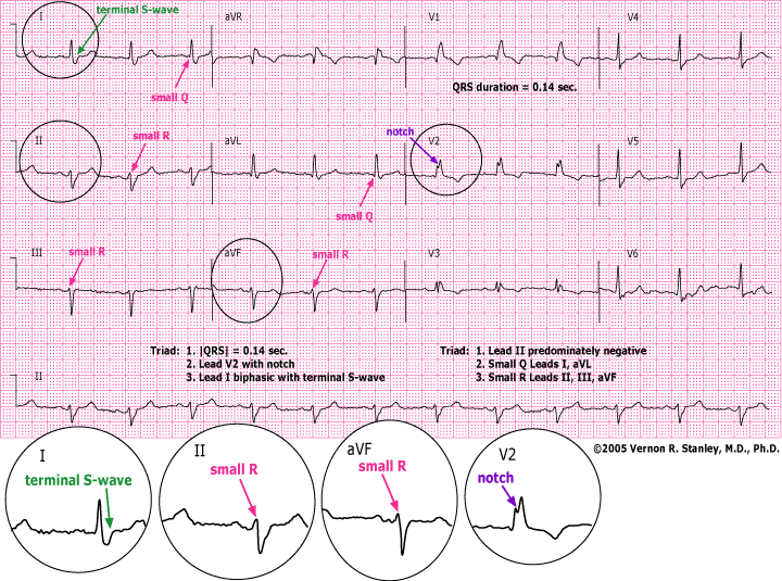
As we interpret this ECG above, we conclude the following:
- There is a Bifascicular Block consisting of the RBBB and LAFB
- ST-T wave changes are present and secondary to the RBBB pattern
- A predominately negative Lead II will determine that the electrical axis lies between – 30 and – 210 degrees
PRIMARY & SECONDARY T-WAVE CHANGES OF THE LBBB & RBBB
SECONDARY T-WAVE CHANGES (in all leads): T-wave is upright if terminal portion of QRS complex is negative. T-wave is inverted if terminal portion of QRS complex is positive.
PRIMARY T-WAVE CHANGES (in all leads): If the above T-wave polarity rule is violated, it is described as primary T-wave changes. This is consistent with myocardial ischemia.
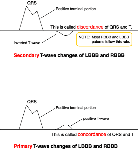
If you find an LBBB (Left Bundle Branch Block) or RBBB (Right Bundle Branch Block), you must further analyze all the T-Waves to categorize them as either Primary or Secondary.
If Primary T-Waves are found, this is consistent with ischemia.
- If you find an LBBB, look for Primary and Secondary T’s. (If primary T-wave changes are present, consider myocardial ischemia.)
- If you find an RBBB, look for Primary and Secondary T’s. (Analyze all the Leads looking for ST-Elevation, ST-Depression, T-Wave Peaking and Significant Q’s that might suggest acute ischemia or infarction.)
Answer key: Q1. “B. Lead I” | Q2 “B. Secondary…” | Q3 “A. True”
Questions about this post or our ECG courses? Contact Us
-
12-lead ECG Interpretation Textbook
$64.99 -
12-lead ECG Interpretation Workbook
$48.50 -
12-lead Textbook, Workbook & HEART Ruler Packet
$124.00Original price was: $124.00.$110.00Current price is: $110.00.


5 Responses
Great review regarding RBBB and LBBB.
Thank you! So glad you enjoyed it!
Thank you for a quick but important review of concordant and discordant ST segments ! Hoping this will stick firmly in my brain 🙂
You are most welcome! I was just speaking with Dr. Stanley about this blog; and he said in his residency and early ER days, the distortions that RBBB and LBBB had on the T-waves and ST segments was always challenging. Kindly, the attending cardiologist took time to educate him, and now we hope to pay it forward! Appreciate your participation! Courtney